
Introduction
Disclosing the complexities of biological regulatory mechanisms and metabolic pathways at the molecular, cellular, tissue, and systemic levels continues to be a central challenge in contemporary biomedical research, particularly in efforts to elucidate the origins and development of cancer. Alongside these scientific efforts, there is an increasing focus on designing alternative in vitro approaches for toxicological assessment of chemical compounds, motivated by both ethical considerations and the need for more efficient research models. A significant barrier in this area is the conception of robust non-animal testing strategies that can reliably anticipate the potential adverse effects of substances following repeated systemic exposure. For such substitutive methods to be effective, it is essential to account for the physicochemical characteristics of the tested compounds, including their aggregation state and pharmaceutical formulation [1, 2].
Among the most intensively studied classes of compounds are copper-based coordination complexes (CCCs), which have attracted substantial attention due to the versatile redox behavior and biological compatibility of copper ions. The pharmacological profile of these metal complexes can be precisely modulated by altering the nature of the coordinating ligands and donor atoms. Particularly, copper complexes have shown considerable promise as antitumor agents, providing a potentially less toxic and more accessible alternative to traditional chemotherapeutics. In addition, their therapeutic applications extend to the management of inflammatory conditions and infectious diseases, including tuberculosis, malaria, and fungal infections [3, 4].
The antitumor efficacy of copper complexes is largely explained by their ability to induce intracellular accumulation of reactive oxygen species (ROS), a process facilitated by their oxidation-reduction potential. The resultant enhancement in ROS levels stimulates cellular antioxidant defense systems in response to oxidative stress (OS). These results highlight the therapeutic potential of ROS-generating copper complexes as antiproliferative agents in cancer treatment strategies [5, 6].
Cellular homeostasis depends critically on balanced redox signaling, where ROS function as physiological mediators. Disruption of this equilibrium contributes to multifactorial pathologies, with malignancies exhibiting particularly pronounced dysregulation. Cancer cells generate elevated ROS through accelerated metabolic activity ‒ a double-edged phenomenon that both triggers tumor progression and gives rise to targetable vulnerabilities for therapeutic exploitation. Selective cytotoxicity can be achieved by further OS elevation in these compromised cells. OS emerges from imbalance between ROS production and antioxidant capacity, causing cumulative damage to proteins, lipids, and DNA. The molecular deterioration accelerates aging processes and initiates carcinogenic pathways. Malignancies exhibit amplified redox discrepancy due to combined endogenous/exogenous reactive species (ROS/RNS), which disrupt signaling networks and promote oncogenic transformation [7].
To counteract such risks, cells rely on a sophisticated network of antioxidant defense mechanisms, encompassing both enzymatic and non-enzymatic components. Key markers of this system include total antioxidant activity (TAA, often measured by ABTS assay), total antioxidant capacity (TAC), and antioxidant substance mass (ASM). The antioxidants, distributed across various cellular compartments, function in unison to ensure reliable protection against oxidative assaults [8, 9].
The complex duality of ROS involved in both tumor initiation and therapeutic targets highlights the central role of oxidative stress modulation in oncology. CCCs represent potential therapeutic agents in the treatment of multifactorial diseases due to their ability to regulate antioxidant systems [10]. This research examines the in vitro antioxidant properties of biologically active thiosemicarbazones using human-derived biological specimens.
The aim of this study was to evaluate the biological potential of copper-based coordination compounds with different thiosemicarbazones by examining their influence on antioxidant markers in vitro in the supernatant obtained from the peripheral blood of clinically healthy donors.
Material and methods
Study design and biological sample preparation. The research was conducted in vitro using biological samples collected in accordance with current principles of biological standardization for experimental procedures. The study protocol was approved by the Research Ethics Committee of the Nicolae Testemițanu State University of Medicine and Pharmacy of the Republic of Moldova (approval no. 5, ref. no. 38, dated June 20, 2024). The study was conducted in compliance with the Declaration of Helsinki and its subsequent amendments (Somerset West Amendment, 1996), regarding the use of human subjects in research [11]. Enrollment in the study was contingent upon participants providing written informed consent.
The research focused on copper-based coordination compounds featuring thiosemicarbazone ligands, which were synthesized at the Advanced Materials Research Laboratory in Biopharmaceutics, Moldova State University [12].
Substances used in the in vitro experiment. The test compounds were classified into the following distinct categories:
Benzothiazole derivatives of thiosemicarbazone – CMA-18, CMD-8, MG-22;
Phenyl thiosemicarbazone derivatives – CMC-34, CMJ-33, CMT-67;
Allyl thiosemicarbazone derivatives – CMG-41, TIA -123, TIA-160.
Table 1. Copper-based coordination compounds with thiosemicarbazones included in the research [13]. | ||
No. | Code | Chemical name of the substance |
1 | Control | 0.1 ml of 0.9% saline solution + Dulbecco’s modified eagle medium (DMEM) |
2 | DOXO | Doxorubicin |
Benzothiazole derivatives of thiosemicarbazone | ||
3 | CMA-18 | Chloro-{1-(1,2-benzothiazol-3-yl)-2-[1-(pyridin-2-yl)ethylidene]diazanido}copper |
4 | CMD-8 | Chloro-{4-ethyl-2-[phenyl(pyridin-2-yl)methylidene]hydrazine-1-carbothioamido} copper |
5 | MG-22 | Chloro-{N'-(4-methoxyphenyl)-N,N-dimethylcarbamimidothioato} copper |
Phenyl thiosemicarbazone derivatives | ||
6 | CMC-34 | Chloro-{N'-[phenyl(pyridin-2-yl)methylidene]-N-pyridin-2-ylcarbamohydrazonothioato} copper |
7 | CMJ-33 | Chloro-{4-(3-methoxyphenyl)-2-[1-(pyridin-2-yl)ethylidene]hydrazine-1-carbothioamido} copper |
8 | CMT-67 | Nitrato-{N-phenyl-N'-(pyridin-2-ylmethylidene)carbamohydrazonothioato} copper |
Allyl thiosemicarbazone derivatives | ||
9 | CMG-41 | Nitrato-{N'-[phenyl(pyridin-2-yl)methylidene]-N-prop-2-en-1-ylcarbamohydrazonothioato} copper |
10 | TIA-123 | Chloro-{N'-[phenyl(pyridin-2-yl)methylidene]-N-prop-2-en-1-ylcarbamohydrazonothioato} copper |
11 | TIA-160 | Acetato-{2-({[(methylsulfanhyl)(prop-2-en-1-lamino)ethylidene]hydrazinylidene} methyl) enolato}copper |
Note: Code – identifier assigned to each compound by the synthesizing researcher; DOXO – Doxorubicin (positive control); Compounds are categorized based on the structure of their thiosemicarbazone ligand: benzothiazole, phenyl, or allyl derivatives. All chemical names refer to copper-based coordination compounds used in the in vitro assays. | ||
Experimental design. Peripheral blood specimens were obtained from 10 healthy volunteers to assess the bioactivity of local copper-based coordination compounds (CCCs). Samples were collected via cubital venipuncture (5 mL/subject) in the morning under fasting conditions using adapted vacutainers.
Sample preparation. Blood was immediately transferred to sterile culture flasks containing 20 mL Dulbecco’s modified eagle medium (DMEM) supplemented with:
Heparin (2.5 IU/mL)
Gentamicin (100 µg/mL)
L-Glutamine (0.6 mg/mL)
Aliquots (0.9 mL) of this suspension were distributed into 24-well plates for antioxidant response assessment.
Treatment protocol. Control wells (n = 4) received 0.1 mL 0.9% NaCl. The other wells were supplemented with the studied CCCs (CMA-18, CMD-8, MG-22, CMC-34, CMJ-33, CMT-67, CMG-41, TIA-123, TIA-160) at final concentrations of 10.0 µM/L and 1.0 µM/L. The compounds were diluted in physiological saline (0.1 mL/well). All experimental treatments were performed twice to ensure data reproducibility.
Incubation and processing. Plates underwent 48-hour incubation at 37°C/3.5% CO2. Post-incubation, well contents were:
transferred to 2.0 mL Eppendorf tubes;
centrifuged (3000 rpm, 5 min);
supernatants aliquoted into 1.5 mL cryovials;
stored at −40°C until analysis.
Blinded coding ensured sample anonymity and traceability throughout the study.
Evaluation of antioxidant system markers. All biochemical assays used to evaluate the antioxidant system parameters were carried out in the Biochemistry Laboratory of Nicolae Testemițanu State University of Medicine and Pharmacy of the Republic of Moldova. The methods originally described by Re R. et al. and Zhang M. et al., were adapted and optimized for application with the Synergy H1 Hybrid Microplate Reader (BioTek Instruments, USA) [14, 15].
In the collected supernatant, the following antioxidant system markers were assessed using spectrophotometric micromethods: total antioxidant activity (TAA using ABTS), total antioxidant capacity (TAC), antioxidant substance mass (ASM), antioxidant total activity (ATA). All parameters were expressed in conventional units (c.u.).
The determination of total antioxidant activity using ABTS (TAA-ABTS) was performed according to the method described by Re R. [14], with modifications introduced by Gudumac V. et al. [16]. This assay is based on the neutralization of the 2,2'-azino-bis-3-ethylbenzothiazoline-6-sulfonic acid radicals (ABTS*) upon interaction with antioxidant compounds present in the biological sample. The neutralization of the radical was measured spectrophotometrically at 734 nm.
The TAC, ASM, and ATA were evaluated based on the method originally described by Zhang M. [15], with adaptations by Tagadiuc O. [17]. This redox-based assay utilizes potassium permanganate (KMnO₄) in the assessment of all the mentioned parameters in biological samples. The antioxidants in the sample, inhibit color development in proportion to their concentration. To determine TAC, ASM, and ATA, absorbance was measured initially (A₀) and after 30 minutes of incubation (A₃₀) under continuous agitation (700 rpm) at 570 nm, using serial dilutions of the biological sample in 0.9% saline (1:10; 1:20; 1:40; 1:80; 1:160), followed by the addition of KMnO₄ solution. Dilution factors were logarithmically transformed into the following values: 1.0, 1.3, 1.6, 1.9, and 2.2. Specific formulas were applied to calculate TAC, ASM, and ATA.
The evaluation of the in vitro effects of the CCCs was performed according to the standard methodology described by Ryzhikova S. L. et al. [18].
Statistical data processing. Data analysis was performed using SPSS v23 (IBM Corp., Armonk, NY). After verifying data distribution and homogeneity of variances, intergroup differences in biochemical parameters were assessed via one-way ANOVA with Games-Howell post-hoc testing for multiple comparisons. Statistical significance was established at p < 0.05. Results are reported as medians with interquartile ranges (IQR).
Results
The present study aimed to investigate the antioxidant activity of copper coordination compounds with thiosemicarbazones (CCCTs), bearing various substituents (benzothiazole, phenyl, and allyl groups), tested at two distinct concentrations (10.0 μM/L and 1.0 μM/L). The effects were evaluated in comparison to both the control group and the pharmacological reference standard ‒ doxorubicin. The results of the biochemical analyses are summarized in Figure 1.
The analysis of the results of TAA evaluation confirms that the compounds CMA-18 and CMD-8 produced a statistically significant decrease at both concentrations, ranging from 10% to 24%, p < 0.001, exhibiting moderate pro-oxidant effects, with inhibition of antioxidant activity at both tested concentrations. Simultaneously, in the in vitro testing of the coordination compound MG-22, a non-significant decrease was recorded at both concentrations, ranging from 1% to 6%, p > 0.05, compared to the control group, maintaining almost the entire antioxidant activity. These findings suggest effective oxidative protection and enhanced cellular tolerance.
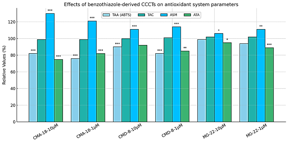 |
Fig. 1 In vitro effects of benzothiazole-derived copper coordination compounds with thiosemicarbazones at concentrations of 10.0 μM/L and 1.0 μM/L on antioxidant system parameters Note: The values for the control group were taken as the reference (100%). Statistical significance comparative to the control group: * − p < 0,05; ** − p < 0,01; *** − p < 0,001; TAA (ABTS): total antioxidant activity measured with ABTS; TAC: total antioxidant capacity; ASM: antioxidant substance mass; ATA: antioxidants total activity. |
The total antioxidant capacity (TAC) of human serum results from the combined contribution of enzymatic and non-enzymatic branches of the system. Oxidative stress arises when the balance between the production of ROS and the body's natural antioxidant mechanisms is disrupted, impairing the efficient neutralization of these reactive species [19].
In conditions where the antioxidant system is functioning suboptimally, the harmful effects of the free radicals become more pronounced. Previous research has predominantly concentrated on evaluating tissue antioxidant capacity, either through the analysis of key antioxidant enzyme activities or by determining levels of low-molecular-weight non-enzymatic antioxidants [20-23]. Therefore, assessing the overall antioxidant capacity of the body has lately become a major area of interest in biomedical research.
The in vitro evaluation of the influence of benzothiazolic copper coordination compounds with thiosemicarbazones on total antioxidant capacity at concentrations of 10.0 μM/L and 1.0 μM/L revealed that all tested compounds maintained or slightly enhanced TAC in a non-significant manner. Notably, MG-22 emerged as the most promising candidate, showing a consistent increase in TAC by 2% at both concentrations (p > 0.05 vs. control group).
In parallel, ASM was significantly elevated under the action of the local compound CMA-18, suggesting a substantial increase in antioxidant load. This may reflect an adaptive response to OS and potential activation of endogenous defense mechanisms. Meanwhile, CMD-8 induced a moderate increase in antioxidant content (p > 0.001), likely associated with mild OS and compensatory activation of antioxidant responses. By contrast, MG-22 induced the lowest increase in antioxidant mass, implying a diminished oxidative risk and a more sensitive cellular activation, with moderate effects on the antioxidant system.
The results of the evaluation of TAA under the influence of CMA-18 revealed a significant decrease at both concentrations, ranging from 18% to 25% (p < 0.001), indicating a pronounced pro-oxidant effect. This reduction in total antioxidant capacity suggests a redox imbalance. At the same time, the compound CMD-8 exhibited a milder effect, although it still led to a slight decrease in TAA, particularly at the lower concentration, with a reduction of 15% (p < 0.01). Conversely, MG-22 proved to be the most promising compound, showing only a minimal decrease in TAA with statistically significant values at both doses. This outcome suggests a favorable antioxidant profile, especially at the higher concentration.
All three compounds studied (CMA-18, CMD-8, and MG-22) demonstrated concentration- and structure-dependent modulation of in vitro antioxidant system parameters (Table 2).
CMA-18 exhibits a distinctly pro-oxidant profile, potentially associated with oxidative cytotoxic effects, as it significantly reduced both TAA and TAC to 75–82% of control values. Although it induced a substantial increase in ASM up to 130%, this appears to represent a compensatory cellular response rather than a protective effect.
CMD-8 demonstrates a moderate oxidative impact, accompanied by mild activation of compensatory antioxidant mechanisms. It is more balanced than CMA-18, causing a slight reduction in TAA and TAC, while inducing a moderate increase in ASM (111–114%). TAC levels remained near control values (100–101%), suggesting preserved systemic antioxidant function.
MG-22 presents the most favorable antioxidant profile, displaying well-regulated redox effects and potential for biomedical applications with low oxidative risk. It maintained TAC and TAA at values close to the control (95–102%) and induced only a minimal increase in ASM, indicating a limited oxidative stimulus and reduced impact on antioxidant defenses.
The evaluation of the TAA of the phenyl group compounds showed that CMC-34 demonstrated a significant decrease in antioxidant activity compared to the control at both tested concentrations (25% –26% reduction, p < 0.001), suggesting a strong inhibitory effect on antioxidant defense mechanisms. In contrast, CMJ-33, at both 10.0 μM/L and 1.0 μM/L, exhibited a slightly more favorable profile, with values of 77.05 μM/L; IQR 3.34; and 78.68 μM/L; IQR 3.28, corresponding to 92% and 94% of the control, respectively. These findings indicate an antioxidant activity nearly comparable to the control group (see Figure 2). CMT-67, at both concentrations (10.0 μM/L and 1.0 μM/L), did not show statistically significant deviations from the control, with values of 82.10 μM/L; IQR 5.12; and 83.28 μM/L; IQR 1.83; representing 98% and 99%, respectively. This outcome reflects a stable antioxidant effect, almost identical to that of the untreated samples.
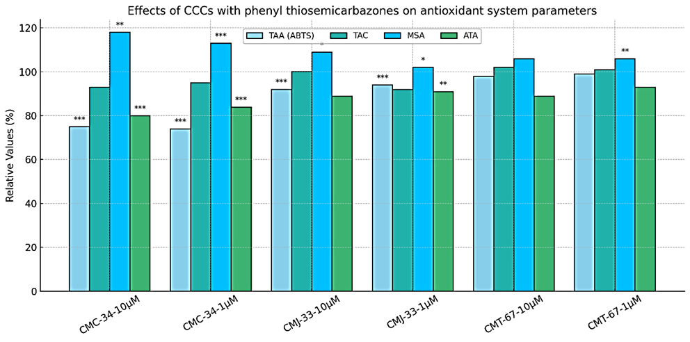 |
Fig. 2 In vitro effects of copper coordination compounds with phenyl thiosemicarbazones at 10.0 μm/l and 1.0 μm/l on antioxidant system parameters Note: The values for the control group were taken as the reference (100%). Statistical significance relative to the control group: * − p< 0,05; ** − p< 0,01; *** − p< 0,001; TAA (ABTS): total antioxidant activity measured with ABTS; TAC: total antioxidant capacity; ASM: antioxidant substance mass; ATA: antioxidants total activity. |
Statistical analysis of TAC for the compound CMC-34 at both concentrations (10.0 μM/L and 1.0 μM/L) showed slightly reduced, non-significant values (5% and 7%). Similarly, CMJ-33 at the concentration of 1.0 μM/L also showed an inconclusive 8% decrease in TAC, while at the concentration of 10.0 μM/L TAC did not change compared to the control (by 0.2%). The compound CMT-67 demonstrated the best results among all tested compounds. At both concentrations of 10.0 μM/L and 1.0 μM/L, TAC values at the control level (+ 1% and 2%), indicating a a neutral and protective effect on the marker values. Therefore, among the phenyl group derivatives, the compound CMT-67 stands out as the most efficient in terms of total antioxidant capacity, with a clear stimulation of cellular defense against free radicals.
Evaluation results of ASM following the action of the phenyl compound CMC-34 showed the most significant increases: 18%, p < 0.01 (at 10.0 μM/L) and 13%, p < 0.001 (at 1.0 μM/L), compared to the control group. This suggests a clear activation of the antioxidant system as a compensatory response to oxidative stress. CMJ-33 induced a modest increase at both concentrations (2–9%), with statistically significant activation at 10.0 μM/L, p < 0.05. Meanwhile, for CMT-67, values remained constant at both concentrations (6% increase), with significance only at 1.0 μM/L, p < 0.01 (Figure 3).
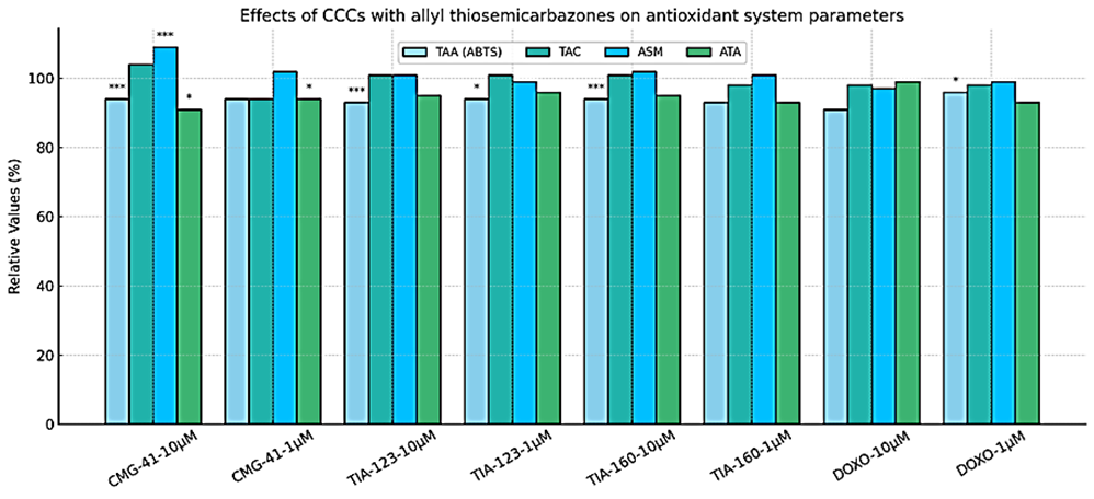 |
Fig. 3 The action of copper coordination compounds with allyl-thiosemicarbazone in vitro at concentrations of 10.0 μm/l and 1.0 μm/l on antioxidant system parameters Note: The values for the control group were taken as the reference (100%). Statistical significance relative to the control group: * − p< 0,05; ** − p< 0,01; *** − p< 0,001; TAA (ABTS): total antioxidant activity measured with ABTS; TAC: total antioxidant capacity; ASM: antioxidant substance mass; ATA: antioxidants total activity. |
Statistical analysis of TAA under the influence of the local compound CMC-34 showed the lowest values at both concentrations (a 16-20% reduction), p < 0.001. These reductions indicate a clear pro-oxidant effect, reflecting redox imbalance. Following CMJ-33 administration, a moderate reduction of total antioxidant activity was observed at both concentrations (a 9–11% reduction), statistically significant only at 1.0 μM/L, p < 0.01. In the case of CMT-67, values remained relatively close to the control: 2.57 and 2.68 c.u. (a 7%–11% reduction). The effect was minimal and without notable statistical significance, indicating good antioxidant stability and maintenance of values close to control at both concentrations.
The three compounds studied (CMC-34, CMJ-33, CMT-67) demonstrated concentration- and structure-dependent modulation of antioxidant system parameters in vitro (Table 2).
CMC-34 exhibited a clear pro-oxidant profile, characterized by a significant decrease in TAA of 16–20% and a slight, non-significant reduction in TAC of 5–7%. Despite this redox imbalance, it triggered a notable enhancement in ASM - up to 18% at 10.0 μM/L - indicating compensatory activation of endogenous antioxidant defenses in response to OS.
CMJ-33 demonstrated a moderate oxidative effect, with a small reduction in TAA (9–11%) and marginal changes in TAC (0–8% decrease), suggesting partial impairment of antioxidant defenses. ASM increased slightly (2–9%), reflecting a modest cellular response to oxidative stimuli, particularly at the higher concentration. Overall, CMJ-33 maintained intermediate redox behavior with limited antioxidant disruption.
CMT-67 displayed the most stable redox profile within the phenyl group derivatives. TAA and TAC remained close to control levels (–7% to –11% and +1% to +2%, respectively), while ASM showed minimal increase (~6%), indicating preservation of redox balance. These findings suggest that CMT-67 exerts a neutral or slightly protective antioxidant effect, making it the most promising candidate among phenyl thiosemicarbazones.
The statistical results of total antioxidant activity (TAA) influenced by CCCs with allyl-thiosemicarbazone, tested in vitro at both concentrations, showed a reduction ranging from 6% to 7%. This decrease was statistically significant at 10.0 μM/L (p < 0.001), while at 1.0 μM/L, significance was observed for TIA-123 (p < 0.05), but not for CMG-41 and TIA-160 (p > 0.05). Additionally, the reference compound doxorubicin, tested in vitro as a positive control, showed the most pronounced reduction in antioxidant activity - by 9% at 10.0 µM/L (p > 0.05) and by 4% at 1.0 μM/L (p < 0.05) compared to the control group, as shown in Figure 3.
TAC values remained generally stable across all compounds, with only minor deviations from the control. Notably, CMG-41 at 10.0 μM/L slightly exceeded the control value by 4%, however at 1.0 μM/L, TAC decreased by 6% (p > 0.05). Other compounds (TIA-123, TIA-160, and DOXO) showed minimal variation, remaining within the 98–101% range relative to the control.
Regarding the antioxidant substance mass (ASM), a statistically significant increase was observed for CMG-41 at 10.0 μM/L (9%, p < 0.001), indicating a potential strong stimulation of antioxidant defense. For TIA-123, ASM remained close to control values with no relevant variation. TIA-160 also maintained nearly identical ASM values at both concentrations (2.71 and 2.70 c.u., ~101–102%), suggesting a consistent antioxidant effect. In contrast, doxorubicin produced a 1% to 3% decrease in ASM at both concentrations (p > 0.05), compared to the control group.
In the assessment of total antioxidant activity (TAA), CMG-41 demonstrated a statistically significant reduction of 6%–9% at both concentrations (p < 0.05). The other tested compounds produced insignificant reductions in TAA, ranging from 4% to 7% (p > 0.05). Similarly, statistical evaluation of doxorubicin revealed a modest decline in TAA of 1%–7% (p > 0.05), indicating a minimal influence on overall antioxidant activity.
The three compounds studied (CMG-41, TIA-123, TIA-160) demonstrated moderate and structure-sensitive antioxidant modulation in vitro.
CMG-41 exhibited a dual behavior, with a statistically significant reduction in TAA (6–9%) and a minor increase in ASM (up to 9%), particularly at 10.0 μM/L, suggesting an adaptive antioxidant response to mild oxidative pressure. TAC showed a slight elevation (+4%) at the higher concentration, indicating possible activation of antioxidant reserves. This profile reflects a mild pro-oxidant potential with compensatory stimulation of defenses.
TIA-123 demonstrated a stable antioxidant effect, with only marginal reductions in TAA (4–5%) and minimal deviations in TAC (98–100%). ASM remained within normal range, reflecting limited oxidative impact. The results indicate a balanced redox profile, with low cellular stress induction and good tolerability across concentrations.
TIA-160 displayed the most favorable antioxidant behavior among allyl derivatives. Both TAA and TAC were well maintained, showing negligible variation from control values (1–2% difference). ASM also remained stable (~101–102%), suggesting excellent redox stability and minimal oxidative burden. This profile highlights TIA-160 as a potentially safe and biocompatible candidate for biomedical use.
Discussion
The present study demonstrates that copper coordination compounds with thiosemicarbazone (CCCTs) exhibit concentration- and structure-dependent redox modulation, disclosing distinct mechanisms of action across three structural classes. Our findings align with emerging research on metal-based therapeutics, particularly regarding their dual capacity to induce OS in malignant cells while preserving redox homeostasis in healthy systems.
Regarding redox profiling and structural correlations, the benzothiazole derivative MG-22 emerged as a lead compound, maintaining TAA and TAC near physiological levels (94–102% of control) while minimally elevating ASM (106–111%). This balanced profile suggests regulated ROS modulation without overwhelming antioxidant defenses, in clear opposition to the pronounced pro-oxidant effects of CMA-18 (TAA reduction to 76–82%). Such divergence underscores how ligand modifications (e.g., methoxy substitution in MG-22) critically determine biological outcomes. These observations resonate with studies showing that N4-methoxyphenyl thiosemicarbazones enhance copper complex stability and redox selectivity [2, 24].
The phenyl derivative CMT-67 displayed TAC conservation (101–102% of control), indicating sustained antioxidant stimulation. This aligns with evidence that CCCTs can activate endogenous antioxidant enzymes like superoxide dismutase (SOD) through copper ion delivery [25, 26]. Conversely, allyl derivatives (e.g., CMG-41) induced ASM spikes (109% increase) alongside a decrease in TAA, making evident adaptive cellular responses to moderate oxidative challenge.
Table 2. Comparative analysis of copper-based coordination compounds with different thiosemicarbazones. | |||||
Compound | TAA (% of Control) | TAC (% of Control) | ATA (% of Control) | ASM (% of Control) | Redox Interpretation |
CMA-18 | ↓ 18–24% (76–82%) | ≈ 99% | ↓ 18–25% (75–82%) | ↑ up to 130% | Strong pro-oxidant effect; cytotoxic potential; compensatory antioxidant response |
CMD-8 | ↓ 10–18% (82–90%) | ≈ 100–101% | ↓ 8–15% (85–92%) | ↑ 111–114% | Moderate oxidative effect; partial antioxidant compensation; balanced redox behavior |
MG-22 | ≈ 94–99% | ↑ up to 102% | ≈ 95–98% | ↑ 106–111% | Favorable redox profile; low oxidative burden; minimal stress response |
CMC-34 | ↓ 16–20% (80–84%) | ↓ 5–7% | ↓ 18–20% (80–82%) | ↑ 13–18% | Pro-oxidant behavior; significant oxidative burden; compensatory antioxidant activation |
CMJ-33 | ↓ 9–11% (89–91%) | ↓ 0–8% | ↓ 9–11% (89–91%) | ↑ 2–9% | Mild oxidative effect; limited stress induction; partial compensatory response |
CMT-67 | ↓ 7–11% (89–93%) | ≈ 101–102% | ↓ 7–11% (89–93%) | ↑ 6% | Stable redox behavior; preserved antioxidant function; minimal oxidative effect |
CMG-41 | ↓ 6–9% (91–94%) | ↑ 4% (at 10 μM/L) | ↓ 6–9% | ↑ 9% | Mild oxidative modulation; adaptive antioxidant stimulation |
TIA-123 | ↓ 4–5% (95–96%) | ≈ 98–100% | ↓ 4–5% | ≈ 100% | Balanced redox profile; negligible oxidative stress; good tolerability |
TIA-160 | ↓ 4–7% (93–96%) | ≈ 101–102% | ↓ 4–7% | ≈ 101–102% | Redox stability maintained; minimal stress response; potentially safe profile |
Note: Data are presented as percentage variation relative to the control group (set as 100%). TAA: Total Antioxidant Activity (assessed via ABTS assay); TAC: Total Antioxidant Capacity; ATA: Antioxidants Total Activity; ASM: Antioxidant Substance Mass. ↑ indicates increase vs. control; ↓ indicates decrease vs. control; ≈ indicates no significant change | |||||
As for therapeutic implications for oncology, the pro-oxidant effects observed in CMA-18 and CMC-34 (TAA reduction to 75–85%) may exploit the "redox vulnerability" of cancer cells, which operate at higher basal ROS levels than healthy cells [24, 26]. This supports the therapeutic strategy of further elevating OS in malignancies to trigger selective cytotoxicity, a concept that was validated by the copper-thiosemicarbazones' ability to induce cuproptosis, a copper-dependent cell death pathway [25, 28]. Notably, MG-22's redox-neutral profile suggests potential as a cytoprotective addition to conventional chemotherapeutics, mitigating treatment-induced oxidative damage.
In order to summarize the mechanistic considerations of our study, the compounds' divergent effects on antioxidant parameters (TAA, TAC, ASM) likely reflect a copper redox cycling, as thiosemicarbazone ligands facilitate Cu(II)/Cu(I) transitions, generating ROS via Fenton-like reactions [27, 29]; an enzyme interaction, as CCCTs may directly modulate SOD/catalase activity or expression, as evidenced by TAC stabilization in CMT-67-treated samples and cellular adaptation, as ASM elevations (e.g., 130% with CMA-18) represent compensatory antioxidant synthesis, consistent with Nrf2 pathway activation observed in other thiosemicarbazone-treated models [26, 30].
This study provides a solid foundation for future research directions but also has several limitations. Although the in vitro model used here provides valuable insights into redox modulation, a key limitation is the lack of testing of the compounds in standardized and pathological cell lines, including tumor-derived cells, limiting the translational significance of the results. Further studies are needed to evaluate the effects of the tested compounds on specific oxidative stress biomarkers and to conduct correlation analyses to establish a comprehensive understanding of their pro- or antioxidant potential. Future studies should also confirm these results in disease-related models, particularly those assessing tumor growth inhibition. Additionally, evaluation of CCCTs in combination with ROS-enhancing therapies such as radiotherapy and in-depth investigation of copper chelation dynamics using advanced techniques such as X-ray absorption spectroscopy are needed to fully elucidate the therapeutic potential of these novel native compounds.
CCCTs represent a promising class of redox modulators with tunable biological effects. Structural optimization (e.g., benzothiazole with methoxy groups) yields compounds like MG-22 that maintain redox equilibrium, while pro-oxidant variants (CMA-18) offer therapeutic potential through selective oxidative cytotoxicity. These findings underscore copper-thiosemicarbazones' versatility in targeting oxidative stress pathways for oncological applications.
Conclusions
Copper coordination compounds with thiosemicarbazone demonstrate significant biomedical potential owing to their redox-active properties and multifunctional bioactivities, including antitumor, antimicrobial, and antioxidant effects. These compounds exhibit complex modulation of antioxidant pathways, dynamically influencing the equilibrium between reactive oxygen species (ROS) generation and cellular antioxidant defenses.
In vitro investigations reveal specific thiosemicarbazones alter antioxidative defense markers: total antioxidant activity; total antioxidant capacity; mass of antioxidant substances; antioxidant total activity, impairing cellular ROS-neutralizing capacity. Such modulation suggests therapeutic utility in pathologies driven by oxidative stress, including degenerative disorders, inflammatory conditions, and multifactorial diseases.
Competing interests
None declared.
Authors’ contributions
VP conceived the study, designed the study, analyzed the data, drafted the manuscript. EP participated in study design, statistical analysis and helped drafting the manuscript. SS critically revised the manuscript and analyzed the data. OT, VG obtained project funding, critically evaluated the results and assessed their applicability. AG offer the analyzed compounds and critically revised the manuscript. All authors reviewed the work critically and approved the final version of the manuscript.
Informed consent for publication
Obtained.
Acknowledgements and funding
The research was funded by the Government of the Republic of Moldova, Ministry of Education and Research, under the institutional subprogram Non-communicable diseases – prevention, diagnosis and personalized treatment (Code 080101, implementation period 2024-2027).
Ethics approval
The study protocol was approved by the Research Ethics Committee of the Nicolae Testemițanu State University of Medicine and Pharmacy, under approval no. 5, ref. no. 38, dated June 20, 2024. The study was conducted in compliance with the Declaration of Helsinki and its subsequent amendments, regarding the use of human subjects in research.
Provenance and peer review
Not commissioned, externally peer review.
Authors’ ORCID IDs
Valeriana Pantea – https://orcid.org/0000-0002-8835-6612
Ecaterina Pavlovschi – https://orcid.org/0000-0003-0385-4805
Stratulat Silvia – https://orcid.org/0000-0003-0985-307X
Aurelian Gulea – https://orcid.org/0000-0003-2010-7959
Olga Tagadiuc – https://orcid.org/0000-0002-5503-8052
Valentin Gudumac – https://orcid.org/0000-0001-9773-1878
References
Pantea V, Cobzac V, Tagadiuc O, Palarie V, Gudumac V. In vitro evaluation of the cytotoxic potential of thiosemicarbazide coordinating compounds in hepatocyte cell culture. Biomedicines. 2023;11(2):366. https://doi.org/10.3390/biomedicines11020366.
Shakya B, Yadav PN. Thiosemicarbazones as potent anticancer agents and their modes of action. Mini Rev Med Chem. 2020;20(8):638-661. doi: 10.2174/1389557519666191029130310.
Krasnovskaya O, Naumov A, Guk D, Gorelkin P, Erofeev A, et. al. Copper coordination compounds as biologically active agents. Int J Mol Sci. 2020;21(11):3965. doi: 10.3390/ijms21113965.
Neguta E, Balan G, Gulea A, et al. Antimicrobial and antifungal activity of Cu(II) and Bi(III) complexes based on amino-polycarboxylate ions and 2-formyl and 2-acetylpyridine thiosemicarbazones. One Health Risk Manag [Internet]. 2021;2(4S):52 [cited 2025 Apr 12]. Available from: https://journal.ohrm.bba.md/index.php/journal-ohrm-bba-md/article/view/….
Pantea V, Andronache L, Globa P, Pavlovschi E, Gulea A, Tagadiuc O, Gudumac V. Copper coordination compounds with thiosemicarbazones: In vitro assessment of their potential
in inhibiting glioma viability and proliferation. Arch Balk Med Union. 2023;58(3):234-244. https://doi.org/10.31688/ABMU.2023.58.3.02.Kim SJ, Kim HS, Seo YR. Understanding of ROS-inducing strategy in anticancer therapy. Oxid Med Cell Longev. 2019;2019:5381692. doi: 10.1155/2019/5381692.
Trapali M, Pavlidis V, Karkalousos P. Molecular insights into oxidative stress and its clinical implications. Open Med Chem J. 2025;19:e18741045373435. http://dx.doi.org/10.2174/0118741045373435250415115811.
Zalewska-Ziob M, Adamek B, Kasperczyk J, Romuk E, Hudziec E, et. al. Activity of antioxidant enzymes in the tumor and adjacent noncancerous tissues of non-small-cell lung cancer. Oxid Med Cell Longev. 2019;2019:2901840. doi: 10.1155/2019/2901840.
Rusu ME, Fizeșan I, Vlase L, Popa DS. Antioxidants in age-related diseases and anti-aging strategies. Antioxidants (Basel). 2022;11(10):1868. doi: 10.3390/antiox11101868.
Gorrini C, Harris IS, Mak TW. Modulation of oxidative stress as an anticancer strategy. Nat Rev Drug Discov. 2013;12(12):931-47. doi: 0.1038/nrd4002.
Puri KS, Suresh KR, Gogtay NJ, Thatte UM. Declaration of Helsinki, 2008: implications for stakeholders in research. J Postgrad Med. 2009;55(2):131-4. doi: 10.4103/0022-3859.52846.
Gulea A, Poirier D, Roy J, Stavila V, Bulimestru I, et. al. In vitro antileukemia, antibacterial and antifungal activities of some 3d metal complexes: chemical synthesis and structure - activity relationships. J Enzyme Inhib Med Chem. 2008;23(6):806-18. https://doi:10.1080/14756360701743002.
Pantea V. Efectele metabolice ale compuşilor biologic activi autohtoni cu acţiune antitumorală [The metabolic effects of native bioactive compounds with antitumor activity] [dissertation]. Chisinau: Nicolae Testemițanu State University of Medicine and Pharmacy; 2023. 128 p. Romanian.
Re R, Pellegrini N, Proteggente A, Pannala A, Yang M, Rice-Evans C. Antioxidant activity applying an improved ABTS radical cation decolorization assay. Free Radic Biol Med. 1999;26 (9-10):1231-7. doi: 10.1016/s0891-5849(98)00315-3.
Zhang M, Liu N, Liu H. Determination of the total mass of antioxidant substances and antioxidant capacity per unit mass in serum using redox titration. Bioinorg Chem Appl. 2014;2014:928595. doi: 10.1155/2014/928595.
Gudumac V, Rîvneac V. Tagadiuc O, et al. Metode de cercetare a metabolismului hepatic: Elaborare metodică [Methods of investigating liver metabolism: Methodological guidelines] Chişinău; 2012. 162 p. Romanian.
Tagadiuc O, Andronache L, Pantea V, Gudumac V, Șveț I, Sardari V. Metodă pentru determinarea capacității antioxidante totale, masei substanțelor antioxidante și a activității mediia antioxidanților în probele biologice [Method for determining total antioxidant capacity, mass of antioxidant substances and average antioxidant activity in biological samples]. Republic of Moldova Certificate of Innovation no. 5641. 2018 March 26.
Ryzhikova SL, Druzhinina IuG, Riabicheva TG, Varaksin NA. Standartizatsiia metodiki opredeleniia produktsii tsitokinov kletkami krovi ex vivo [The standardization of technique of detection of blood cells cytokine production ex vivo. Klin Lab Diagn. 2011;(11):49-53. Russian.
Hood E, Simone E, Wattamwar P, Dziubla T, Muzykantov V. Nanocarriers for vascular delivery of antioxidants. Nanomedicine (Lond). 2011;6(7):1257-72. doi: 10.2217/nnm.11.92.
Zhang M, Liu N, Liu H. Determination of the total mass of antioxidant substances and antioxidant capacity per unit mass in serum using redox titration. Bioinorg Chem Appl. 2014;2014:928595. doi: 10.1155/2014/928595.
Kattamis C, Lazaropoulou C, Delaporta P, Apostolakou F, Kattamis A, et al. Disturbances of biomarkers of iron and oxidant-antioxidant homeostasis in patients with beta-thalassemia intermedia. Pediatr Endocrinol Rev. 2011;8 Suppl 2:256-62.
Zujko ME, Witkowska AM. Dietary antioxidants and chronic diseases. Antioxidants (Basel). 2023;12(2):362. doi: 10.3390/antiox12020362.
Munteanu IG, Apetrei C. Analytical methods used in determining antioxidant activity: a review. Int J Mol Sci. 2021;22(7):3380. doi: 10.3390/ijms22073380.
Rusnac R, Garbuz O, Kravtsov V, Melnic E, Istrati D, Tsapkov V, Poirier D, Gulea A. Novel copper(II) coordination compounds containing pyridine derivatives of N4-methoxyphenyl-thiosemicarbazones with selective anticancer activity. Molecules. 2024;29(24):6002. doi: 10.3390/molecules29246002.
Vo TTT, Peng TY, Nguyen TH, Bui TNH, Wang CS, Lee WJ, Chen YL, Wu YC, Lee IT. The crosstalk between copper-induced oxidative stress and cuproptosis: a novel potential anticancer paradigm. Cell Commun Signal. 2024;22(1):353. doi: 10.1186/s12964-024-01726-3.
Zilka O, Poon JF, Pratt DA. Radical-trapping antioxidant activity of copper and nickel bis(thiosemicarbazone) complexes underlies their potency as inhibitors of ferroptotic cell death. J Am Chem Soc. 2021;143(45):19043-19057. doi: 10.1021/jacs.1c08254.
Singh NK, Yadav PN, Kumbhar AA, Pokhrel YR. Anticancer potency of copper(II) complexes of thiosemicarbazones. J Inorg Biochem. 2020:111134. doi: 10.1016/j.jinorgbio.2020.111134.
Luo M, Zhou L, Huang Z, Li B, Nice EC, Xu J, Huang C. Antioxidant therapy in cancer: rationale and progress. Antioxidants (Basel). 2022;11(6):1128. doi: 10.3390/antiox11061128.
Kowol CR, Heffeter P, Miklos W, Gille L, Trondl R, Cappellacci L, Berger W, Keppler BK. Mechanisms underlying reductant-induced reactive oxygen species formation by anticancer copper(II) compounds. J Biol Inorg Chem. 2012;17(3):409-23. doi: 10.1007/s00775-011-0864-x.
Leal M, Silva M, Marques D, Mendes R, Ximenes R, Machado D, Silva Jj, Rodrigues C, Cruz F, Lima M. Preliminary evaluation of the toxicological, antioxidant and antitumor activities promoted by the compounds 2,4-dihydroxybenzylidene-thiosemicarbazones an in silico, in vitro and in vivo study. An Acad Bras Cienc. 2024;96(2):e20231247. doi: 10.1590/0001-3765202420231247.